What is De Quervain Disease?
The tendons enabling the movements of the thumb is preserved by a surrounding sheath at the level of wrist (fig.1) the strain and stretching of the tendons cause to reaction and edema (fluid retention on the tissues). This situation results in the strain and decrease of volume in the sheath. The pain increases and it gets into a vicious circle. We can call these conditions as “inflammation”.
The most important reason of the strain and irritation of the tendons at the first stage is to repeatedly make a new movement that the wrist is not used to. Especially the mothers who has newly given a birth are under this risk. Holding the baby continuously and with different positions is a new movement for the wrist and the thumb. Besides, the hormonal changes occurring due to pregnancy and breast-feeding may also affect this process.
The studies show that some people are more prone to de Quervain disease. This means that while you may not observe the disease in every people of the group equally making the same straining movements. De Quervain disease is more frequently seen in the people having anatomic variation (congenital variation) in the sheath preserving the tendons in the wrist. In this text, variation means that the sheath has another pocket (septa) in it or the number of the tendons is more than the average population.
HOW TO DIAGNOSE?
The most significant symptom is the pain on the thumb side of the wrist. It might occur either by slowly increasing or suddenly. The pain may spread to the thumb or up to the arm. The pain may increase with the movements of the hand or the thumb. Especially the wrist movements with a strong grasp (like lifting a teapot) may cause to sudden and severe pain. Swelling, liquid retention feeling and cyst (ganglion) formation on the soft tissue may be observed on the same area (Fig.2)
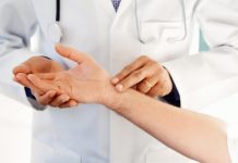
Finkelstein test usually helps to establish the diagnosis (Fig.3). Although it is controversial that wrist joint radiography is required for every patient, it enables not to skip over any additional pathologies. The most frequently seen pathologies are;
1-corrosiion- arthritis on the thumb root (carpometacarpal arthritis)
2- scaphoid ( a small bone on the wrist) fracture.
The conditions like trigger finger, carpal tunnel syndrome and tennis elbow that you will see in the following chapters are the pathologies frequently accompanying to the de quervain disease.
WHAT IS THE TREATMENT?
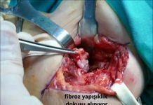
The instruments named splint can be used in order to keep the thumb still. Although it generally helps to avoid the pain at the early stage, the pain recurs at the 70% of the patients. The large majority of the studies shows that the corticosteroid (cortisone) injection (needle) duly applied to the pain area is therapeutical. It is observed that supporting the injection with the splint does not change the result. This injection can safely be applied during pregnancy and breast-feeding period. The success rate is around 80% and success rate of the splint treatment is around 15%; meaning that approximately 4 of every 5 patient can be successfully treated with cortisone injection. Surgical treatment is the only option for the patients having unsuccessful results. During the surgery, the thickened sheath preserving the tendons and the inside pockets (septa) are cut by carefully protecting the adjacent nerve tissues and the tendons are released (Fig. 4,5,6,7,8).
THE PROCESS OF SURGICAL TREATMENT; WHAT IS AHEAD OF US?
The orthopedician or Hand Surgeon may ask for x-ray after the consultation. In the large majority of the surgical intervention, day-case (short term of hospitalization) is applied. There is no need for general anesthesia. Local (axillary block or RIVA) anesthesia is generally sufficient. It is highly important to mention the special conditions (chronic diseases, regularly used medication etc.) during the consultation with your hand surgeon. In the postop. early stage (the first 3 days) cold administration and holding the hand over the heart level will decrease the pain and throbbing. In the early stage, you will have bandage around your wrist. This bandage is generally opened between 5-7 days and the wound is controlled. If no problem is detected, you will be able to take shower. Physical therapy and rehabilitation is rarely needed at the end of this stage. Even though this period may change according to the implemented surgery and the condition of the wrist, it is expected to get back to the routine life within 3 weeks.
POSSIBLE REVERSIONS
The most significant reversion is to injure or cut a nerve (sensory limb of the radial nerve) running though the surgery implemented area (Fig.9). the operation should be performed with special glasses. In case of an injury of the nerve, a long and difficult process begins.
Another possible reversion is that the sheath is not opened sufficiently and the pockets (septa) are not noticed. In this case, the surgery process should be repeated. Congestion (hemotoma) on the operated wound area, infection (inflammation) formation, limitation of the finger movements due to tissue adhesion on the surgery implemented area, chronic pain and getting the results late or never are the most common reversions.
It is important to preserve the nerve (sensory limb of the radial nerve) running through the surgery implemented area.
Prof. Dr. Hakan Gündeş’s Click here for publication on the subject.


