Киста Ганглиона (Рис. 1)
Карпометакарпальный Бугорок (Рис. 9)
Узлы Возникающие в Результате Укола Инородного Предмета (Рис. 10)
[auto_thumb width=”350″ height=”300″ link=”https://www.hakangundes.com.tr/wp-content/uploads/iyi-huylu-tumor1.jpg” lightbox=”true” align=”left” title=”” alt=”” iframe=”false” frame=”true” crop=”true”]https://www.hakangundes.com.tr/wp-content/uploads/iyi-huylu-tumor1.jpg[/auto_thumb]
[auto_thumb width=”350″ height=”300″ link=”https://www.hakangundes.com.tr/wp-content/uploads/karpometalkarpal-boss1.jpg” lightbox=”true” align=”left” title=”” alt=”” iframe=”false” frame=”true” crop=”true”]https://www.hakangundes.com.tr/wp-content/uploads/karpometalkarpal-boss1.jpg[/auto_thumb]
[auto_thumb width=”350″ height=”300″ link=”https://www.hakangundes.com.tr/wp-content/uploads/yabanci-cisim-batmasi1.jpg” lightbox=”true” align=”left” title=”” alt=”” iframe=”false” frame=”true” crop=”true”]https://www.hakangundes.com.tr/wp-content/uploads/yabanci-cisim-batmasi1.jpg[/auto_thumb]
Опухоли образовывающиеся жировыми клетками (Рис. 11, 12)
[auto_thumb width=”350″ height=”199″ link=”https://www.hakangundes.com.tr/wp-content/uploads/yag-hucre-tumorleri.png” lightbox=”true” align=”left” title=”” alt=”” iframe=”false” frame=”true” crop=”true”]https://www.hakangundes.com.tr/wp-content/uploads/yag-hucre-tumorleri.png[/auto_thumb]
[auto_thumb width=”350″ height=”199″ link=”https://www.hakangundes.com.tr/wp-content/uploads/yag-hucre-tumorleri.png” lightbox=”true” align=”left” title=”” alt=”” iframe=”false” frame=”true” crop=”true”]https://www.hakangundes.com.tr/wp-content/uploads/yag-hucresi-tumor.png[/auto_thumb]
Образование мягкой ткани на кисти необходимо убирать под увеличительным прибором
Нейрофибролипома: Жировик состоящий из нервной ткани (нерв пальца)
Гигантоклеточная Опухоль Сухожильного Влагалища (Рис. 13, 14)
[auto_thumb width=”350″ height=”199″ link=”https://www.hakangundes.com.tr/wp-content/uploads/tendon-kilif1.jpg” lightbox=”true” align=”left” title=”” alt=”” iframe=”false” frame=”true” crop=”true”]https://www.hakangundes.com.tr/wp-content/uploads/tendon-kilif1.jpg[/auto_thumb]
[auto_thumb width=”350″ height=”199″ link=”https://www.hakangundes.com.tr/wp-content/uploads/tendon-kilifi1.jpg” lightbox=”true” align=”left” title=”” alt=”” iframe=”false” frame=”true” crop=”true”]https://www.hakangundes.com.tr/wp-content/uploads/tendon-kilifi1.jpg[/auto_thumb]
Рецидив. Гигантоклеточная опухоль сухожильного влагалища. По причине вовлечения капсула сухожилия и сухожильное влагалище удалены.
Figure 14. The Archive of Hakan Gündeş Recurrence of Giant cell tumor of the tendon sheath. The joint capsule and the tendon sheath is remoced due to the involvement.
Гигантоклеточная опухоль сухожильного влагалища. Вовлечение сосудисто-нервного пучка в результате позднего обращения к специалисту и устранение образования после операции. Не смотря на то, что большинство этих опухолей являются доброкачественными, однако есть высокий риск рецидива (повторение опухоли в том же месте).
Шваннома, это опухоль мягкой ткани образующаяся из сухожильного влагалища. Операцию необходимо проводить очень внимательно по причине вероятности рецидива и неврологических повреждений.
[auto_thumb width=”350″ height=”199″ link=”https://www.hakangundes.com.tr/wp-content/uploads/sinir-dokusu.jpg” lightbox=”true” align=”left” title=”” alt=”” iframe=”false” frame=”true” crop=”true”]https://www.hakangundes.com.tr/wp-content/uploads/sinir-dokusu1.jpg[/auto_thumb]
[auto_thumb width=”350″ height=”199″ link=”https://www.hakangundes.com.tr/wp-content/uploads/nurofibrom1.jpg” lightbox=”true” align=”left” title=”” alt=”” iframe=”false” frame=”true” crop=”true”]https://www.hakangundes.com.tr/wp-content/uploads/nurofibrom1.jpg[/auto_thumb]
Фиброма и Нейрофиброма. Доброкачественные опухоли с низкой вероятностью рецидива.
Фиброматозные Опухоли (Рис. 17, 18, 19)
[auto_thumb width=”350″ height=”300″ link=”https://www.hakangundes.com.tr/wp-content/uploads/fibramatoz1.jpg” lightbox=”true” align=”left” title=”” alt=”” iframe=”false” frame=”true” crop=”true”]https://www.hakangundes.com.tr/wp-content/uploads/fibramatoz1.jpg[/auto_thumb]
[auto_thumb width=”350″ height=”300″ link=”https://www.hakangundes.com.tr/wp-content/uploads/fibra-tumor1.jpg” lightbox=”true” align=”left” title=”” alt=”” iframe=”false” frame=”true” crop=”true”]https://www.hakangundes.com.tr/wp-content/uploads/fibra-tumor1.jpg[/auto_thumb]
[auto_thumb width=”350″ height=”300″ link=”https://www.hakangundes.com.tr/wp-content/uploads/fibramatoz-tumorler1.jpg” lightbox=”true” align=”left” title=”” alt=”” iframe=”false” frame=”true” crop=”true”]https://www.hakangundes.com.tr/wp-content/uploads/fibramatoz-tumorler1.jpg[/auto_thumb]
Figure 17. The Archive of Hakan Gündeş Fibromatous tumors Ulnar nerve-vein involvement on the hypothenar area.
Рис.18. Архив Хакан Гюндеша
Figure 19. The Archive of Hakan Gündeş Mass on the thumb root diagnosed with Fibromyositis only after the excisional biopsy.
Фиброматозная опухоль.
Вовлечение локтевого нерва/артерии в гипотенарной области. После удаления образования.
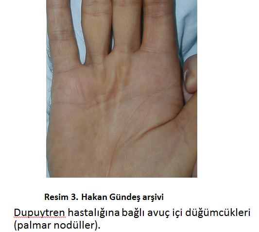
Abnormal Palmar Thickening (Dupuytren, Noldules Fasciitis) (figure 3)
[auto_thumb width=”300″ height=”190″ link=”https://www.hakangundes.com.tr/wp-content/uploads/avuc-ici-kalinlasmasi1.jpg” lightbox=”true” align=”center” title=”” alt=”” iframe=”false” frame=”true” crop=”true”]https://www.hakangundes.com.tr/wp-content/uploads/avuc-ici-kalinlasmasi1.jpg[/auto_thumb]
Аномальное Утолщение в Ладони (Болезнь Дюпюитрена, Узловой Фасциит) (Рис.3)


